Researchers at Hokkaido University are developing a new hyperspectral imaging (HSI) method to detect and quantify cryogenic minerals – particularly gypsum – in sea ice. This innovative approach could help scientists understand how these microscopic crystals influence ocean carbon cycling and the changing polar environment.
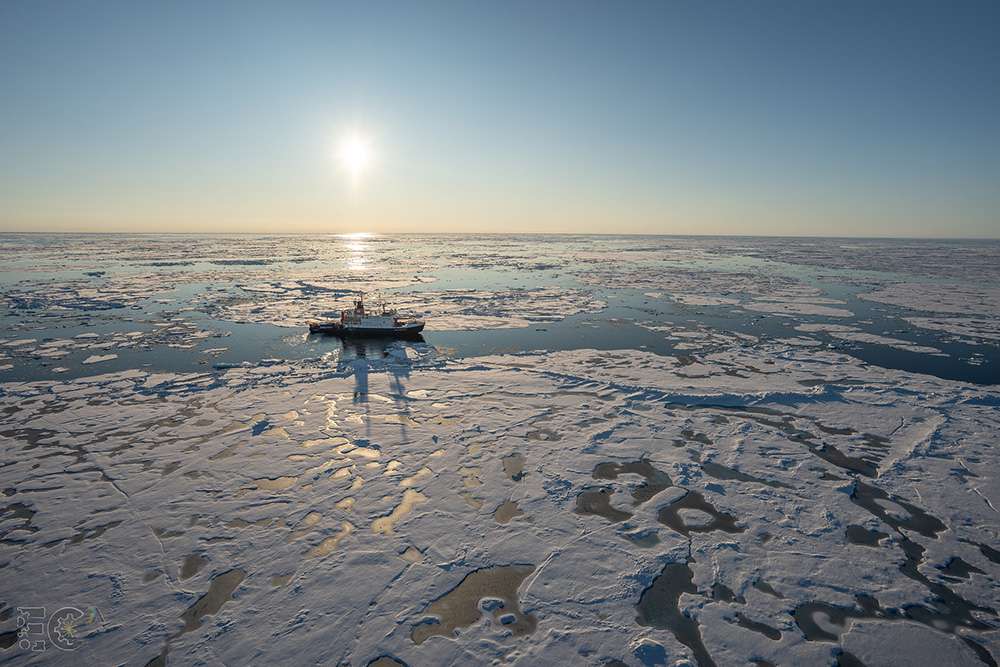
Image 1: Research icebreaker RV Polarstern during the CONTRASTS PS149 expedition. Image courtesy of Emiliano Cimoli.
Revealing what the eye can’t see
Detecting a microscopic crystal trapped inside a larger crystal of ice is a challenge that even advanced microscopes struggle to solve. This problem is evident especially in the polar fieldwork where time and sample volume are limited. While expert input can help identify minerals, traditional manual methods can be slow, subjective, and labour-intensive, posing challenges for automation and making it difficult to quantify mineral abundance across different ice types.
To overcome these limitations, researchers at Hokkaido University, supported by collaboration with the Alfred Wegener Institute (AWI) and the renowned Arctic research capabilities of the RV Polarstern, are testing hyperspectral imaging (HSI) as a faster, and objective method. Using the distinct spectral fingerprints of cryogenic minerals in the near- and shortwave infrared (NIR/SWIR) range, HSI can identify and quantify minerals in sea ice efficiently and non-destructively.
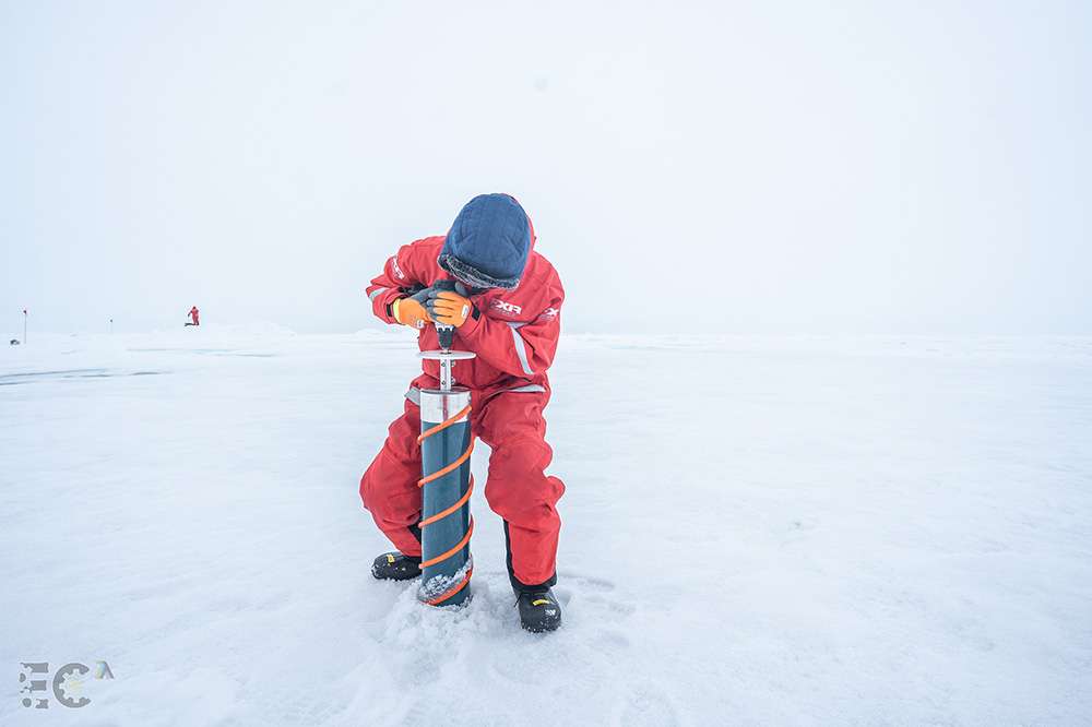
Image 2: Drilling sea-ice core samples. Image courtesy of Emiliano Cimoli
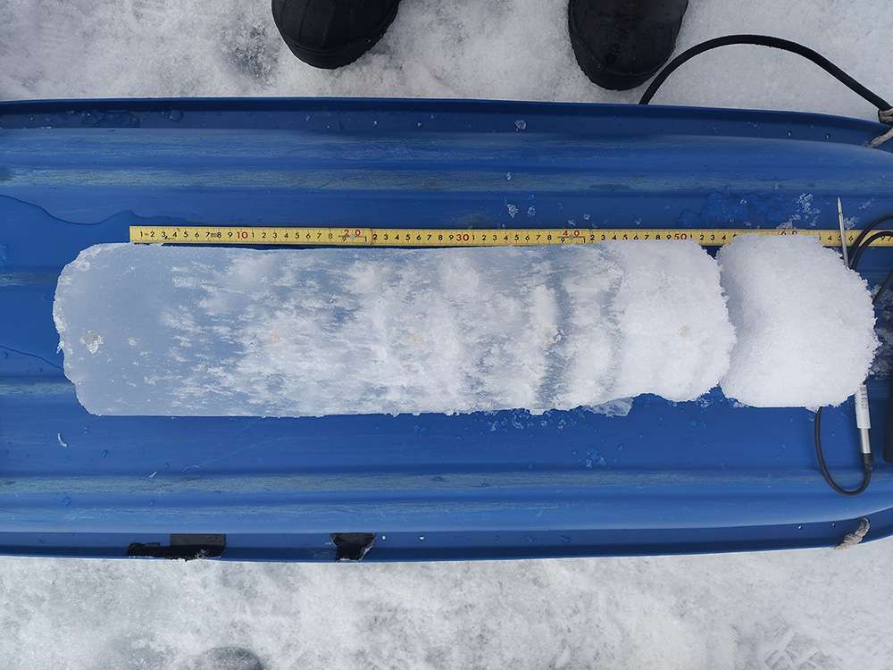
Image 3: Sea-ice core sample. Image courtesy of Emiliano Cimoli
Gypsum: an overlooked mineral with a big role in the ocean
Sea ice is a dynamic, multiphase system filled with tiny brine microenvironments where cryogenic minerals form, often ranging from less than 10 μm to over 1000 μm in size. One such mineral, gypsum (calcium sulfate, CaSO₄·2H₂O), has long been overlooked in polar research despite its potential importance. It may act as a “ballasting agent,” helping organic material sink from the ocean surface to deeper layers – an essential process in the global carbon cycle (see here for further information).
However, gypsum dissolves easily during sampling and transport, meaning it has historically remained under observed. As a result, its ecological significance and distribution remain largely unknown.
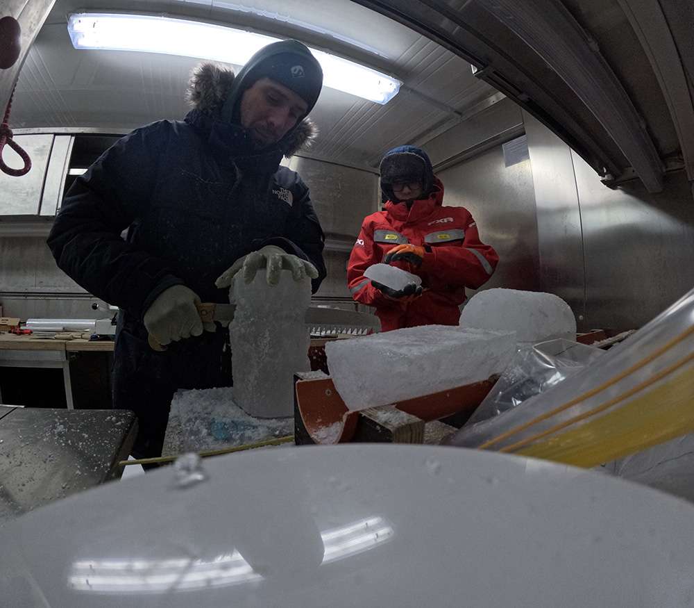
Image 4: Dr. Emiliano Cimoli and Dr. Keigo D. Takahashi processing sea-ice core samples. Image courtesy of Emiliano Cimoli
Adapting geological imaging to polar research
Dr. Emiliano Cimoli, JSPS Research Fellow at the School of Fisheries Sciences, Hokkaido University, leads the effort to apply novel optical sensing techniques to study cryogenic biogeochemical processes in polar environments. He works alongside Dr. Keigo D. Takahashi, also a JSPS Research Fellow, supported by the Japan Society for the Promotion of Science (JSPS) International Postdoctoral Fellowship.
“Drawing inspiration from geological and planetary remote sensing of hydrated sulfates, we aim to adapt these approaches to polar biogeochemical research,” Dr. Cimoli explains.
The team’s goal is to use HSI to detect and quantify gypsum and related cryogenic minerals directly from filters that capture melted sea-ice core samples.
This method enables near real-time mineral detection in the field, allowing semi-automated analysis across different ice types, seasons, and regions.
Finding efficient methods is becoming increasingly important as sea-ice conditions undergo rapid change.
“Hyperspectral imaging can open the door to automated mineral identification, crystal counting, and potentially even size and thickness estimation,” says Dr. Cimoli.
“This throughput-friendly approach could dramatically speed up sample processing and help standardize polar research workflows directly on polar research vessels.”
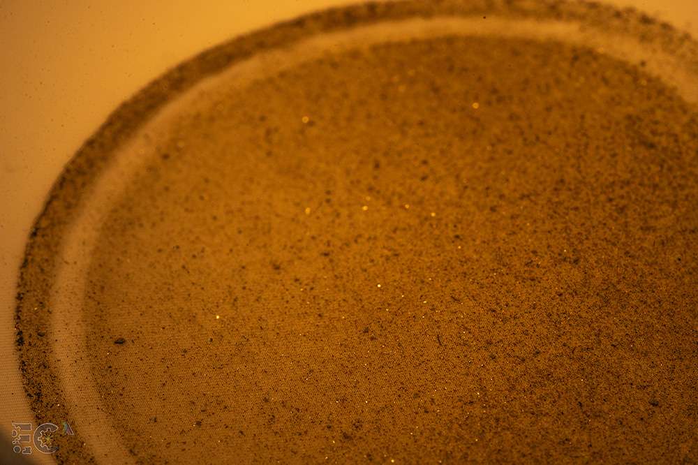
Image 5: Filtered ice sample with potential mineral presence. Image courtesy of Emiliano Cimoli
Specim FX17: seeing what the human eye cannot
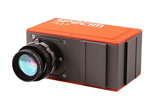
The Specim FX17 hyperspectral camera identifies minerals by capturing their unique absorption features between 900 and 1700 nm. Operating in the near-infrared range, it allows researchers to detect material properties invisible to standard optical instruments.
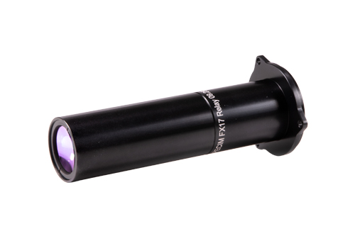
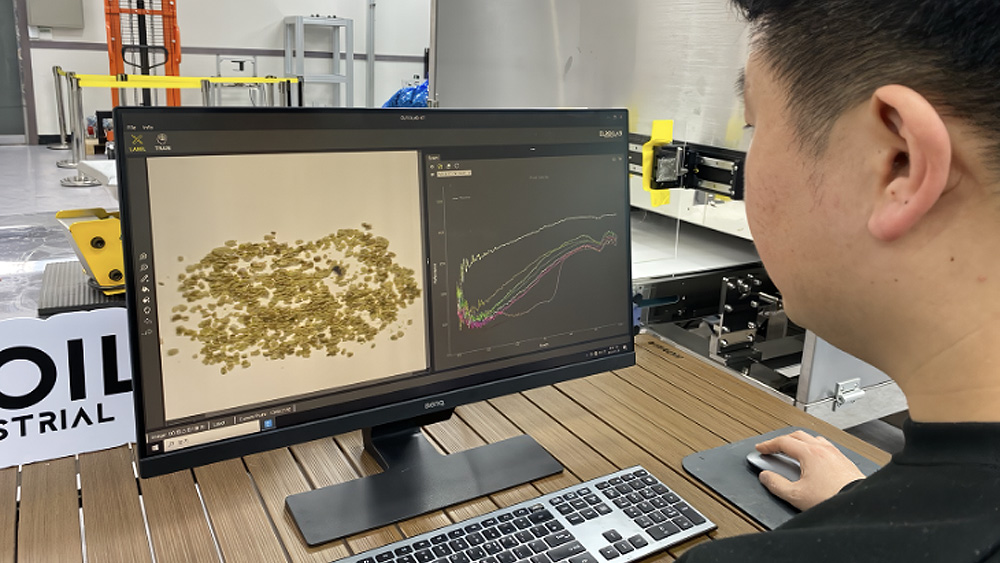
Cimoli and his team use the Specim FX17 with a macro lens setup (approx. 10 μm resolution) and controlled lighting to test a field-adaptable workflow for scanning filters from melted sea-ice cores and quantifying mineral content.
“In our ongoing trials, we melt sectioned sea-ice cores under controlled conditions and filter the remaining material through a 10 µm mesh. We then apply imaging spectroscopy to see whether spectral signatures associated with gypsum and other cryominerals can be reliably captured on these filters,” Cimoli explains.
By successfully estimating mineral presence and distribution, the researchers aim to link these patterns to broader sea-ice properties such as temperature history, porosity, and seasonal stage.
“The Specim FX17 is central to testing whether a rapid, non-destructive, and field-ready workflow can be developed for identifying these elusive mineral phases in polar environments,” Cimoli emphasizes.
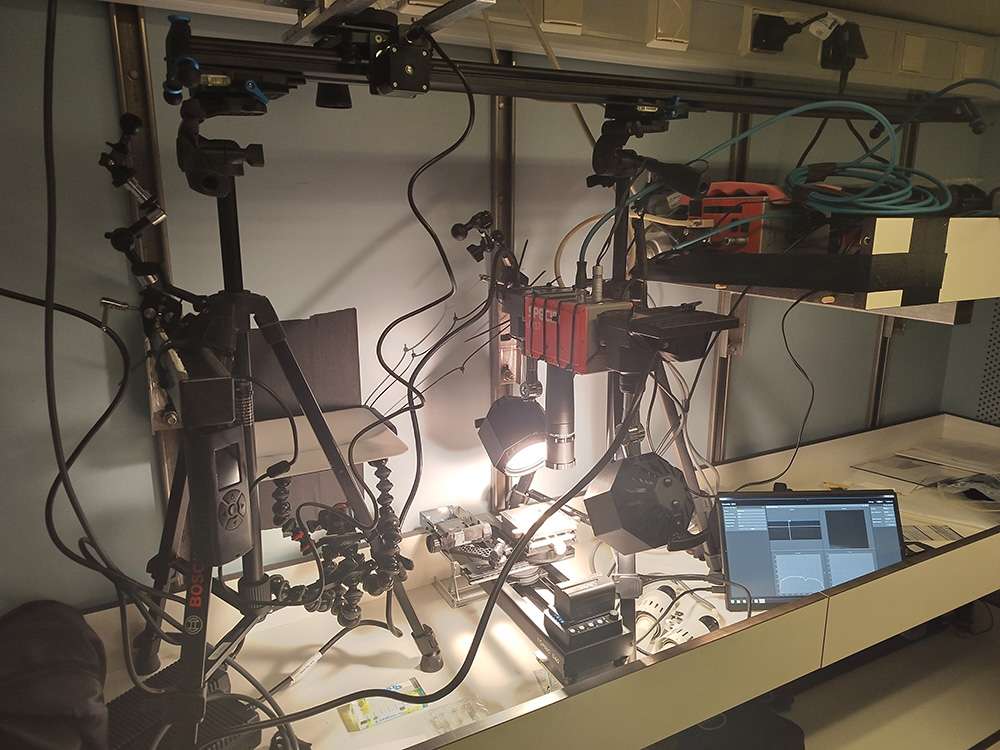
Image 6: The Specim FX17 workstation equipped with the macro lens. Image courtesy of Emiliano Cimoli
A reliable choice for challenging research environments
This is not Cimoli’s first encounter with Specim technology. During his PhD, he used the Specim AisaKESTREL10 for under-ice imaging of sea-ice algal communities – another challenging optical task in extreme environments (Learn more).
Beyond the technical capabilities, he highlights Specim’s reliability and responsive support as crucial for field-based science and the key reasons he continues to rely on Specim products.
“Over the years, I’ve found Specim to be one of the few companies willing to support unusual or experimental applications in demanding conditions,” Dr. Cimoli says.
“Their flexibility and fast response have been invaluable for research that depends on equipment reliability in the field.”
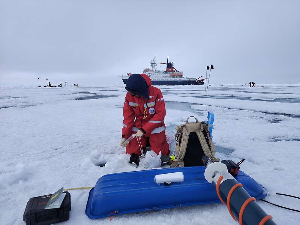
Image 7: Dr. Keigo D. Takahashi immersed in field work on the sea ice. Image courtesy of Emiliano Cimoli
Testing the workflow aboard the RV Polarstern
The Hokkaido University team tested their HSI workflow aboard the German research icebreaker RV Polarstern during the CONTRASTS PS149 expedition, led by AWI. This multidisciplinary mission investigated how atmospheric and oceanic variability influence Arctic sea-ice melt and it associated ecosystems.
The project brought together Japanese, German, and Australian research teams, giving Dr. Cimoli’s group a unique chance to validate their HSI method under real polar conditions. Working closely with AWI’s broader sea-ice physics and biology program helped integrate optical techniques into an interdisciplinary understanding of Arctic change.
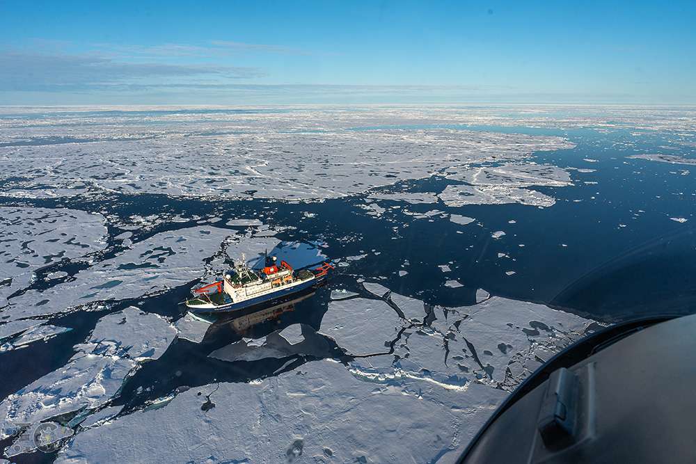
Image 8: RV Polarstern. Image courtesy of Hokkaido University
A new lens on polar science
By combining advanced imaging technology with field-ready workflows, the Hokkaido University project demonstrates how hyperspectral imaging can reveal details of sea-ice composition previously hidden from view.
The approach offers a promising path toward rapid, non-destructive, and standardized mineral detection, helping scientists better understand the evolving polar ecosystem. As sea-ice conditions change faster than ever before, such innovations are essential to capture the full picture of our planet’s frozen frontiers.
A timelapse video of the Specim FX17 workstation in action. Video courtesy of Hokkaido University.
Learn more:
Wollenburg, J. E., Iversen, M., Katlein, C., Krumpen, T., Nicolaus, M., Castellani, G., Peeken, I., and Flores, H.: New observations of the distribution, morphology and dissolution dynamics of cryogenic gypsum in the Arctic Ocean, The Cryosphere, 14, 1795–1808, https://doi.org/10.5194/tc-14-1795-2020
Cimoli, E., Lucieer, V., Meiners, K.M. et al. Mapping the in situ microspatial distribution of ice algal biomass through hyperspectral imaging of sea-ice cores. Sci Rep 10, 21848 (2020). https://doi.org/10.1038/s41598-020-79084-6